|
|
|
Pigments:
Pigments are coloured substances, some of which are
normal constituents of cells (eg melanin), whereas others are abnornmal
and collect in cells onlyu under special circumstances. Pigments can be
either exogenous - coming from outside the body, ot endogenous - synthesised
within the body itself.
Exogenous pigments:
The most common exogenous pigment is cardon, which is
a frequent air pollutant of urban life. When inhaled, it is picked up
by macrophages within alveoli and then trasnpiorted via lymphatics to
regional lymph nodes. Accumulation of this pigemnt in the lung causes
a black discolouration and is termend anthracosis.
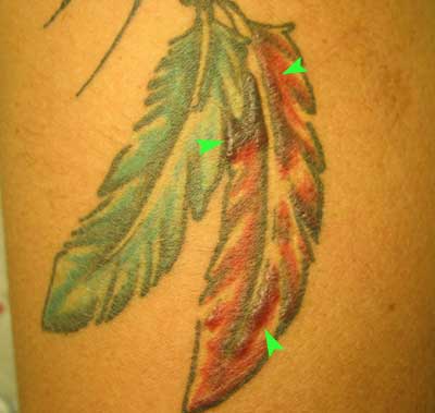
Tattooing is a form of localised exogenous pigmentation
of the skin. The pigments innoculated are generally phagocytosed by dermal
macrophages, in which they reside from then on. The pigments do not usualy
evoke any inflammatroy response.

This young adult developed a granulomatous reaction
(arrowheads) in the red component of a tattoo on the right arm. The tattoo
was acquired a year earlier. Delayed hypersensitivity reactions occur
rarely in tattoos and are most commonly associated with the red dye cinnabar
which contains mercuric sulfide. Reactions have also been reported with
chrome green and cobalt blue.
Endogenous Pigments:
The major endogenous pigemts seen in tissues are: lipofuscin,
melanin, haemosidrin and bilirubin.
Lipofuscin is an insoluble pigment sometimes
referred to as the "wear and tear" pigment. It is thought that
lipofuscin is derived through lipid peroxidation of polyunsaturated lipids
of subcellular membranes. Lipofuscin is not injurious to the cell or its
functions. Its importance lies in its being the telltale sign of free
radical injury and lipid peroxidation. In tissue sections it appears as
a yellow-brown, finely granular intracytoplasmic, often perinuclear pigment.
Melanin is an endogenous, non-haemaglobin derived,
brown-black pigment formed when the enzyme tyrosinase catalyses the oxidation
of tyrosine to dihydroxyphenylalanine in melanocytes.
Haemosiderin is a haemoglobin derived, golden
yellow to brown, granular or crystalline pigment in which form iron is
stored in cells. Iron is nornmally carried by specific transport proteins
- transferrins. In cells, it is stored in association with a protien -
apoferrtin, to form ferritin micelles. Ferritin is a constituent of most
cell types. When there is a local or systemic excess of iron, ferritin
forms haemosiderin granules which are easily seen with the light microscope.
Bilirubin is the major normal pigment found
in bile. It is derived from haemoglobin but contains no iron. Its normal
formation and excretion are vital to health, and jaundice (icterus) is
a common clinical disorder caused by excess of this pigment within cells
and tissues.
 
|

