Introduction
Case
1922/90. Solitary bone swelling in a mature animal
History:
German Shepherd dog aged 7 years. The right distal tibia
became progressively swollen, and the dog became lame. The mass was firm
and on radiology localized bone lysis was evident.
At post mortem there were form pale nodules on the lungs and the tibia
was grossly swollen with multiple irregular necrotic areas within a poorly
circumscribed mass
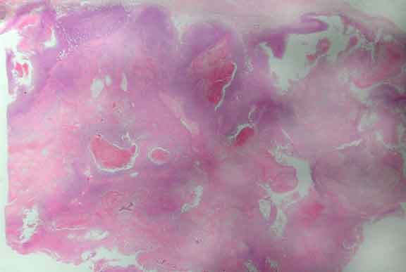
Low power view of lesion
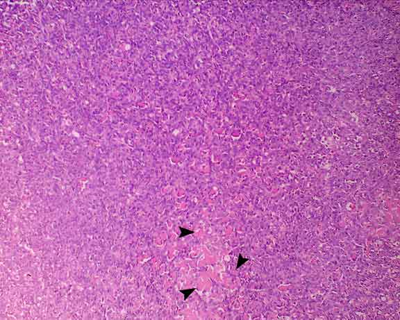
High power view of lesion. Note the pink material (arrowheads) being produced by the neoplastic cells = tumour osteoid - helps to allow classifiation of this spindle cell tumour as an osteosarcoma.
Osteosarcoma in a dog
Concepts to consider:
What are the known causes of bone cancer?
How do bone tumours differ
From those in flat bones
From those in cats
What are the diagnostic features of osteosarcomas?
Could this lesion be confused with
Healing fracture callus?
Osteomyelitis?
Secondary cancer invading bone?