Introduction
Introduction:
One of the basic distinctions that must be made when considering neoplasia is whether the tumour is benign or malignant. Some of the criteria used to make that distinction are outlined in Table 1.
Cases
Review Questions
Back to Prac Classes
Table 1: Criteria used for classification of neoplasms as Benign or Malignant |
||||
Criterion |
Benign |
Malignant |
||
Growth: |
||||
|
slowcircumscribed |
rapidinfiltrative |
||
Mode of Growth: |
||||
|
neverrare |
commoncommon |
||
Cellular Characteristics: |
||||
|
near normalnear normalnear normaluniformnormal, smallfew, normal |
abnormalhyperchromaticenlargedpleomorphiclarge/multiplefrequent, abnormal |
||
Stromal Features: |
||||
|
adequateoften abundantabsentrare |
often scantyvariablecommoncommon |
||
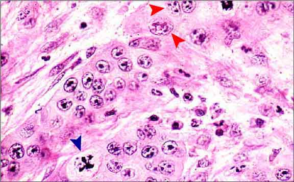
Anaplastic features of a squamous cell carcinoma (= malignant tumour):
-
large central nucleoli (in some cells, multiple nucleoli are present – red arrowheads)
-
Abnormal mitotic figure (blue arrowhead)
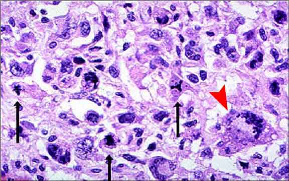
Anaplastic features of a poorly differentiated fibrosarcoma.
Note the atypical mitotic figures (black arrows) and the multinucleate
and uninucleate giant tumour cells (red arrowhead). There is variation
in cell shape and size.

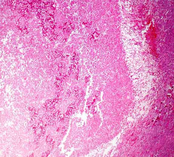
Sertoli cell tumours often incite a large amount of collagen to be deposited by (non-neoplastic) mesenchymal cells. (x40)
Slide Ref 443-03