
Introduction
Case 7:
History:
12 year old Golden Retriever dog with a focal, pedunculated lesion protruding from the rectum.
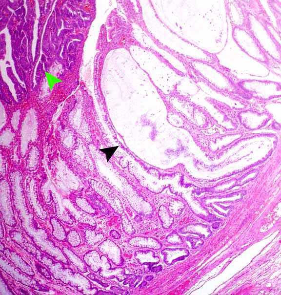
Colon (x40). Segmental transformation of colonic mucosa – neoplastic mucosa is darker blue due to high density of cells (green arrowhead). Non-neoplastic mucosa – some colonic glands have being distorted due to compression by tumour – producing cystic dilation of glands, filled with mucus (black arrowhead).
Slide Ref: 148/03 *
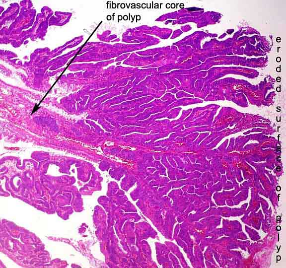
Colon (x40).
Slide Ref: 148/03 *
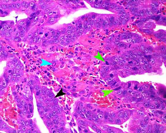
Colon (x400). Pseudostratified cuboidal/columnar cells
sitting on a basement membrane (black arrowhead). Mitotic figures (green
arrowheads).
Inflammatory cells in lamina propria (blue arrowhead)
Slide Ref: 148/03 *