|
|
|
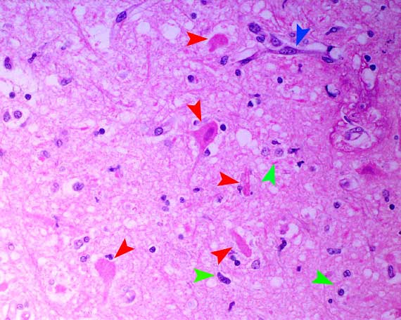
Necrotic neurons - red arrowheads,
gliosis - green arrowheads, reactive endothelium - blue arrowheads.
The appearance of the necrotic neurons is typical of excitotoxic
injury mediated by excessive release of excitatory neurotransmitters
such as glutamate.
Such injury may be seen in cerebral ischaemia, hypoglycaemia,
thiamine deficiency, organic mercurial poisoning, lead poisoning and
in indirect salt poisoning.
In the brain, the neurons that are most vulnerable
to oxygen/energy deprivation lie in certain parts of the cerebral cortex,
cerebellar cortex (especially Purkinje cells), hippocampus, amygdala
and basal and thalamic nuclei and they normally utilise glutamate in
neurotransmission.
Severe seizure activity in humans is well known to
cause secondary neuronal necrosis in the predilection sites in the brain.
Similar lesions have also been noted experimentally in primates and
rodents and observed in seizuring dogs, especially young dogs under
1 year of age.
Hypotension, pyrexia, hypoxia, hypoglycaemia and
neuronal hyperactivity may all contribute to this phenomenon in animals
suffering protracted and severe seizures.
A primary cause of seizure activity was not identified
in this dog. A presumptive diagnosis of idiopathic epilepsy with secondary,
seizure-induced, subacute, hippocampal neuronal necrosis was made.
 
|