|
|
|
Case 4:
History:
A 1.5 year old Thoroughbred colt presented with progressively
deteriorating hindquarter ataxia over an approximately 3 week long period.
The colt was euthanised with suspected cervical stenotic myelopathy (wobbler
syndrome). A small chronic chip fracture of cervical vertebral body 6
was detected at necrcopsy, with palpable softening (malacia) and slight
indentation of the spinal cord at the level of the fracture.
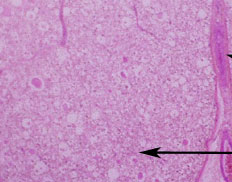
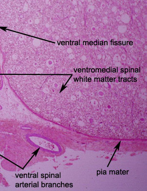
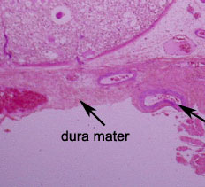
This is a low power view of
the ventromedial aspect of the spinal cord at the level of T3. The
ventromedial location is obvious from the ventral midline fissure.
Central spinal grey matter is not visible. The white matter of the
ventromedial spinal funiculi has a pronounced motheaten appearance
and there are several swollen eosinophilic globular structures.
What are the latter likely to be?
|


