|
|
|
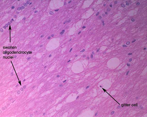
The motheaten appearance of the white matter reflects
a combination of episodes of primary demyelination of initially normal
axons and episodes of concurrent axonal and myelin injury. Occasional
gitter cells within ballooned spaces indicate that there is ongoing disintegration
of axons and myelin.
In distemper infection, white matter lesions are typically
multifocal and especially target tracts close to CSF, e.g. the optic tract,
cerebellar peduncles, the fornix of the hippocampus and spinal white matter.
Productive viral infection occurs in ependymal and choroid plexus epithelial
cells, with virus being disseminated into the CSF and spreading into adjacent
tissue.
|
|