|
|
|

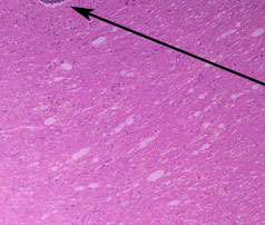
Some of these tight aggregates of chiefly proliferating
microglia are consistent with neuronophagic nodules,
i.e. nodular aggregates of cells phagocytosing dead neurons.
Neuronophagic nodules are often a prominent feature
of viral encephalitides.
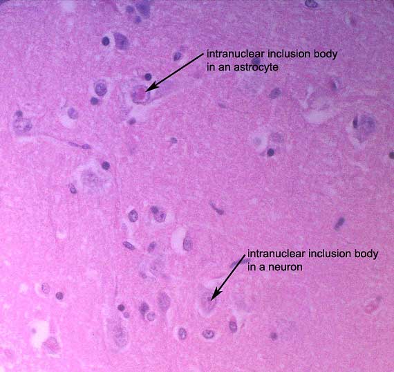
A thorough search of the brain
sections at high power revealed occasional eosinophilic intranuclear
and intracytoplasmic viral inclusion bodies in astrocytes and, less
frequently, neurons.
These inclusion bodies permitted confirmation of a diagnosis of canine
distemper (paramyxoviral) infection.
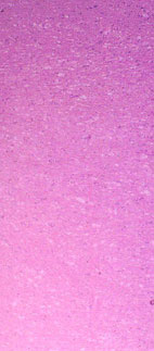
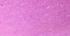
This is a low power view of
a large white matter tract in the brain. The white matter is coarsely
and irregularly vacuolated.
|
|