|
|
|
Case 1:
History:
A 7 week old pit bull terrier pup presented weak, ataxic
and unable to stand. Conjunctivitis and blepharitis were apparent on physical
examination. A littermate had died 2 days previously after a brief illness
characterised by vomiting and terminated by collapse and respiratory arrest.
The pup was treated with antibiotics and intravenous fluids but it
developed dyspnoea and was euthanised. Ante mortem clinical pathology
findings included anaemia (PCV = 0.17 L/L), hypoproteinaemia (44 g/L)
and neutropenia (2.6 x 10^9/L).
Necropsy Findings:
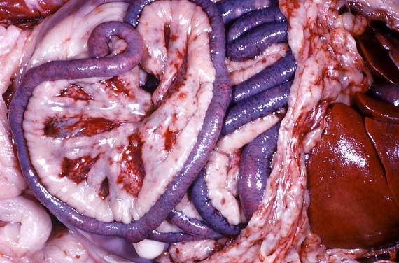
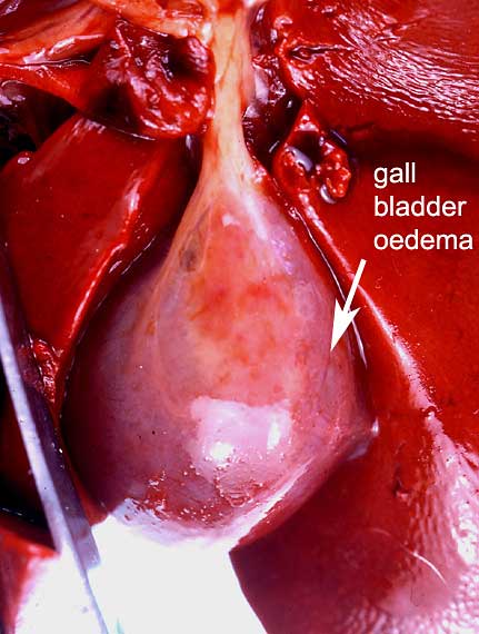
Post mortem examination revealed generalised carcase pallor, a mild
serous peritoneal effusion, petechial and ecchymotic haemorrhages over
the intestines and mesenteries, a few fresh fibrin strands over the liver,
digested blood in the gastric lumen and moderately severe mural oedema
of the gall bladder.
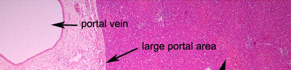
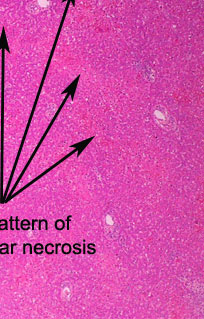
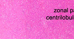
In this low magnification of the liver, a zonal pattern of haemorrhage
and necrosis is apparent (red areas). By identifying the smaller portal
areas, it can be seen that these red areas involve parenchyma distant
from the portal areas (i.e. the centrilobular zones = zone 3).
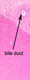
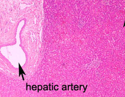
A large portal area lies in the top left of the field. Note the oedematous
distension of the connective tissue of the portal area.
PREVIOUS
|