|
|
|
Case 2:
History:
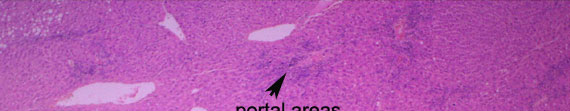
A sheep liver was submitted from an abattoir for histopathology.
The liver was mildly reduced in size and was coarsely multinodular throughout.
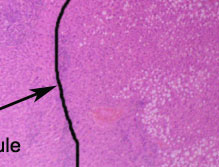
At low magnification, loss of parenchyma (hepatic
atrophy) is suggested by the small size of some of the hepatic
lobules and by the irregular approximation of adjacent portal areas and
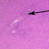
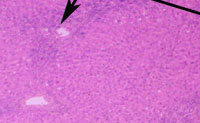
central veins. The portal areas are abnormally prominent and basophilic.
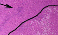
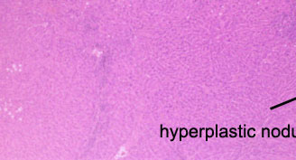
In the lower right corner, there is a large hyperplastic
parenchymal nodule in which the hepatocytes have undergone mild fatty
degeneration.
PREVIOUS
|