Introduction
Case 4:
History:
A 7 year old male Labrador collapsed and died during a round-the-block run. The dog had been clinically normal prior to the collapse.
Necropsy Findings:
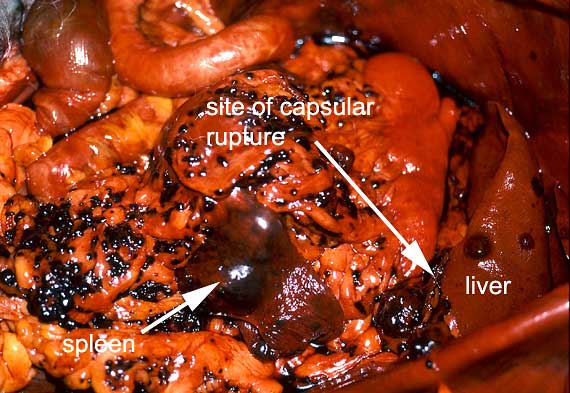
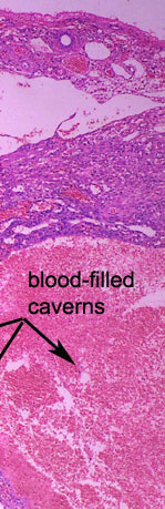
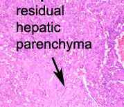
At necropsy, there was approximately 1.8 L of partially clotted blood in the peritoneal cavity. Numerous, small, soft, red-black, blood-filled nodules were widely implanted over the omentum, intestinal mesenteries and right ventral diaphragm. Fewer larger nodules of comparable appearance were present in the spleen and throughout the liver. A blood clot was adherent to the ruptured surface of one such nodule protruding from the visceral aspect of the left medial hepatic lobe, indicating the source of the fatal haemorrhage.
| |
|
|||
 |
 |
 |
|
|
 |
|
|||
 |
 |
|
||
 |
|
|||
 |
|
|||
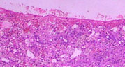
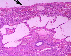
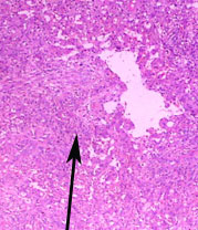
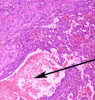
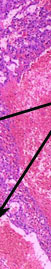
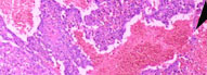
In this low power view of a superficial portion of a liver lobe, there has been partial replacement of the hepatic parenchyma by abnormal tissue forming irregularly shaped and sized blood-filled spaces.