|
|
|
Case 6:
History:
A 6 year old male castrated Siamese cat presented with
a 6 week history of inappetence, weight loss and lethargy. Clinical pathology
abnormalities included a moderately severe non-regenerative anaemia, an
inflammatory leukocytosis, hyperglobulinaemia and mild elevations of serum
ALT, AST, GGT, urea and creatinine.
Necropsy Findings:
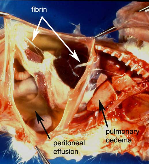
The owners decided upon euthanasia. At necropsy, the cat appeared moderately
dehydrated and there was serous atrophy of body fat stores consistent
with the reported weight loss. A large volume of yellow viscous fluid
with a few strands of fresh fibrin was present in the peritoneal cavity
and a smaller volume of comparable fluid was present in the pleural cavity.
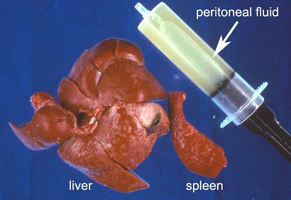
The lungs were heavy with oedema fluid. Numerous miliary, cream-white
foci and small plaques were scattered over the hepatic and splenic capsules,
over the subcapsular parenchyma of the kidneys and over the omentum and
intestinal mesenteries.
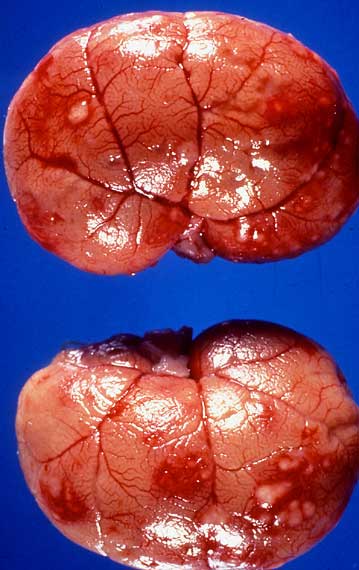
Kidneys
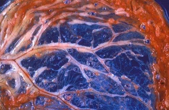
Greater Omentum
Analysis of the peritoneal fluid revealed it to be a
non-septic exudate with a high protein concentration of 73 g/L. Nucleated
cells present comprised sloughed mesothelial cells, non-degenerate neutrophils,
monocytes/macrophages and small lymphocytes. Aerobic and anaerobic cultures
were negative.
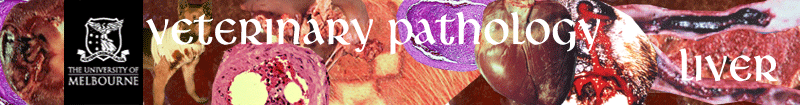
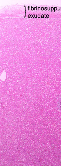
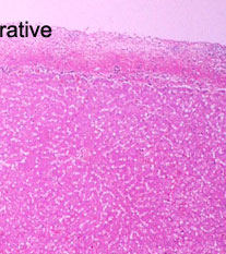
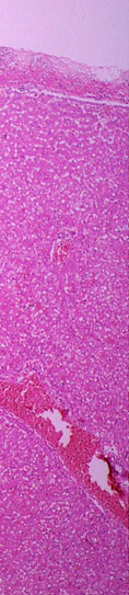
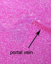
In this low power view of the liver, there is a thick
layer of exudate over the hepatic capsule. In the superficial parenchyma,
a large focus of inflammation and necrosis overlaps a portal vein branch
and extends into the adjacent tissue.
|