|
|
|
Case 2:
History:
-
Arabian filly foal, 6 weeks old
-
Weak foal, respiratory signs from 1 week of age
-
At necropsy, extensive congestion and atelectasis of cranial lung
fields
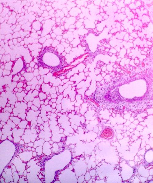
Histopathology shows a disease centred on smaller bronchi
and bronchioles. This is seen as oedema around airways, necrosis of the
airway epitheliam and accumulation of exudate within the airway lumens.
Surrounding lung is only minimally affected.
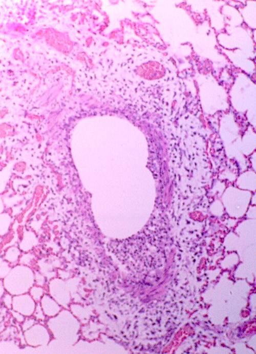 
Examples of airway lesions in a bronchiole (left) and
small bronchus (right).
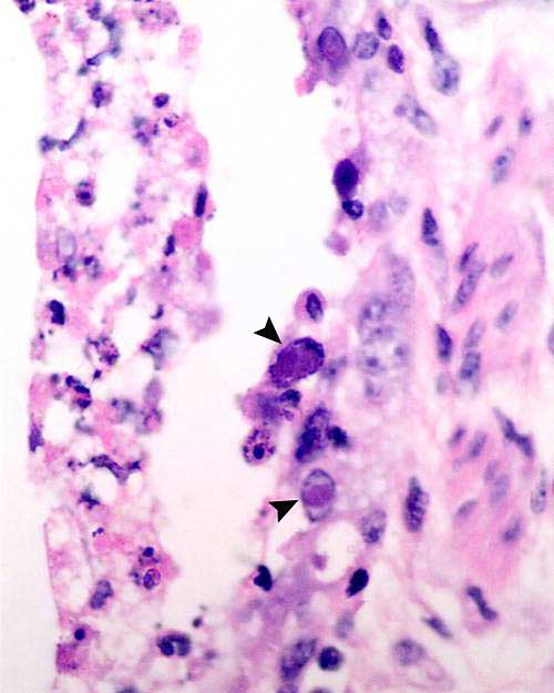
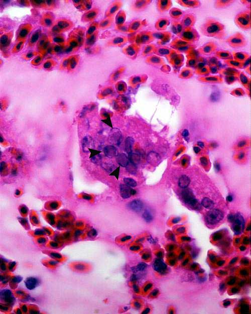
Higher power illustrating necrotic cell debris in the
lumen (left), partially ulcerated mucosa and several epithelial cells
containing large basophilic intranuclear inclusions (arrowheads). The
lesions are typical for adenoviral infection.
Differential Diagnosis for Bronchioloitis:
-
Adenovirus
-
Other viruses eg Herpes
Infectious Laryngotracheitis (ILT) - a classic
herpes virus disease of chickens affecting the airways:
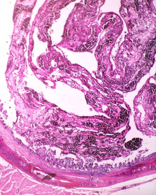
Haemorrhagic to diphtheritic exudates are typical of
Herpes infections of the airways in many species. Another example of a
similar disease is IBR of cattle. The lumen is occluded with bloody exudate
and the mucosa is necrotic and ulcerated.
Often diagnostic inclusions are found in the sloughed
epithelial cells. Severe disease may lead to suffocation. This view shows
amphophilic inclusions (arrowheads) within respiratory epithelial cells
in the exudate in the lumen.
|