|
|
|
Case 2:
History:
Chronic renal failure was diagnosed in a 7 year old
female bull mastiff. She was euthanised due to clinical deterioration
over a 3 week period. At necropsy, both kidneys were misshapen and shrunken.
The pelvis of the left kidney was mildly enlarged and distorted. The bladder
wall was thickened and a film of green-tinged exudate was present over
the mucosal surface.
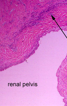
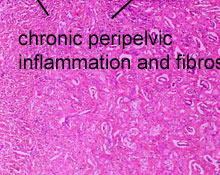
This is a low power view of
the pelvis of the left kidney. Beneath the pelvic epithelium, there
is a thick band of excess collagen which is lightly infiltrated by
mononuclear leukocytes.

|


