|
|
|
Histological Findings:
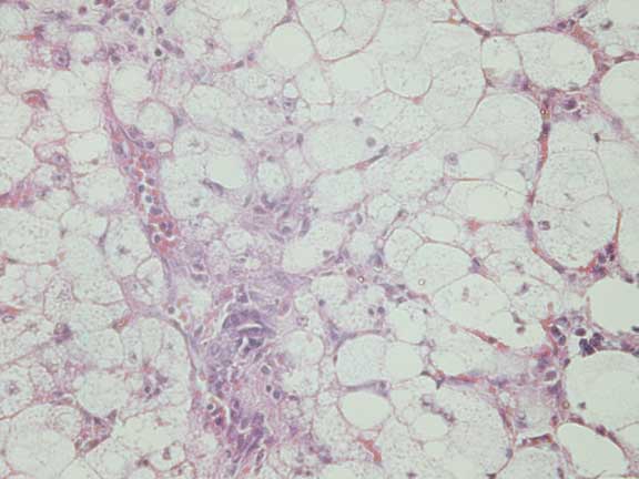
Haematoxylin & Eosin (x400)
At low power, a distinct change can be seen between
the normal hepatocellular architecture and a region of cells that contain
large vacuoles consistent with lipid. The fatty change within the hepatocytes
is uniform across all the acinar zones.
The sharp demarcation, the uniformity of the lesion
and its close proximity to a capsular adhesion all suggest that physical
forces exerted locally to this liver area may have resulted in these changes.
Take a moment to piece together the information presented
and examine the photomicrographs carefully and see if you can determine
how the abnormalities identified may all be linked.
 
|
 Veterinary
Pathology - Cell Degeneration
Veterinary
Pathology - Cell Degeneration