Introduction
Lung:
 |
||
 |
 |
 |
 |
||
Introduction |
Lung:
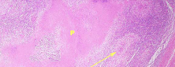
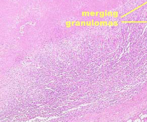
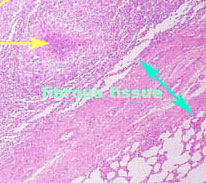
Haematoxylin & Eosin (x100)The lung lesions consisted of a core of amorphous proteinaceous
material (P), which in some cases had become mineralised. Around the core
there was a ring of enlarged activated macrophages with foamy cytoplasm.
Some macrophages showed signs of epithelioid cell formation and a small
number of Langerhans' giant cells are also present. Around the macrophages
are scattered lymphocytes and plasma cells. Fibrous capsules surround
almost all lesions.
|
|||||||||
Cases |
||||||||||
Review Questions |
||||||||||
Back to Prac Classes |
||||||||||