|
|
|
Histological Findings:
 
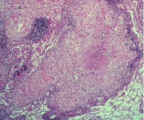
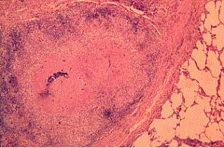
Haematoxylin & Eosin (x40)
The lung tissue between lesions appears relatively normal.
The lesions themselves are discrete nodules which have an eosinophilic
amorphous centre (some containing a basophilic staining granular deposit
consistent with mineral deposition). Surrounding the necrotic centre is
a ring of large macrophage cells and epithelioid giant cells with vacuolated
cytoplasm. Around these are lymphocytes and occasional plasmocytes. Variable
amounts of fibrous tissue are associated with the lesions.
This is an example of what type of necrosis?

|
 Veterinary
Pathology - Necrosis
