|
|
|
Infarction
Infarction = local ischaemic tissue
injury.
Causes include:
-
shock
-
endotoxaemia
-
cardiac failure
-
hypovolaemia
-
obstruction of a blood vessel by an embolus, a thrombus, external
compression etc
Appearance of Infarcts
Gross Appearance
Infarcts may be pale or red. Infarcts are invisible
for the first 12 hours but may thence appear as friable and slightly pale
wedges of tissue. They become progressively highlighted by haemorrhage
and/or by superficial fibrin exudation (if they are covered by a serosal
membrane or capsule). As they age, infarcts become more distinct and paler
than surrounding viable tissue. When chronic and composed of scar tissue,
infarcts are pale, firm and shrunken.
Septic infarcts may be converted into abscesses
if the animal survives.
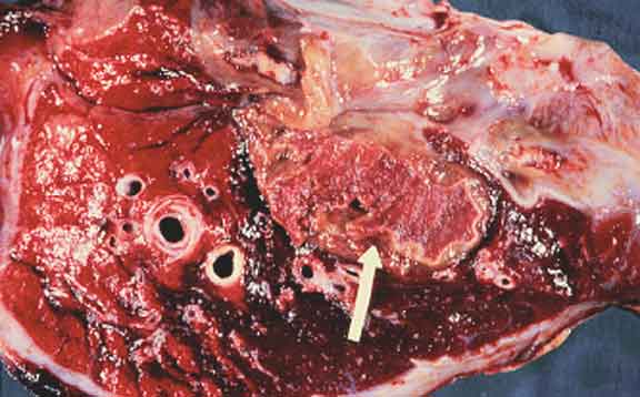
A pulmonary infarct in a horse. How
old do you think this infarct is?
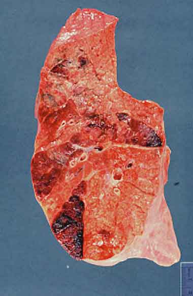
Multifocal haemorrhagic pulmonary infarcts. Note
the typical wedge shape of the infarcts The obstructed vessels would be
located at the apices of the wedges. Pulmonary infarcts are typically
red, due to the spongy nature of the lungs and the presence of a dual
blood supply.
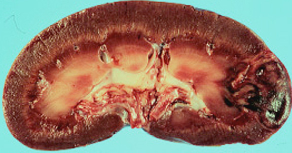
Subacute renal infarct in a dog. Note
the partial dehaemoglobinisation of the centre of the infarct and the
pale band at its margins. The pale band represents the zone of leukocytic
infiltration and early reparative fibroplasia.
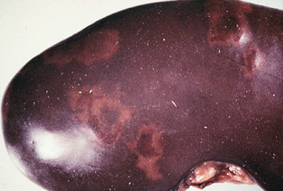
Subacute renal infarcts in a pig.
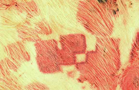
Haemorrhagic cutaneous infarcts in a pig due to acute
Erysipelothrix rhusiopathiae septicaemia (erysipelas).
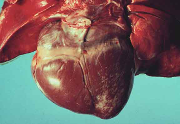
Chronic (scarred) myocardial infarcts in the left ventricle
of a dog. Note that the pale scarred foci are depressed below the surface
of adjacent tissue due to contraction of mature collagen.
Appearance of Infarcts
Histological Appearance
In all tissues except the brain, ischaemic necrosis
is of coagulative type with the mummified remnants persisting for at least
several days as ghost outlines.
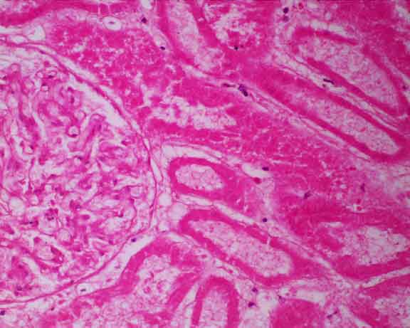
Renal Infarct. Hypereosinophilic remnants of the
renal cortex can still be identified, including profiles of cortical tubules
and a glomerulus. All these elements are dead, as indicated by the nuclear pyknosis or karyolysis. This appearance is typical of ischaemic coagulative
necrosis.
|