| |
|
|
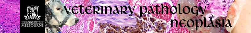
Cardiac mass:
Vascular channels are lined by a continuous layer of
plump endothelial cells. (x200)
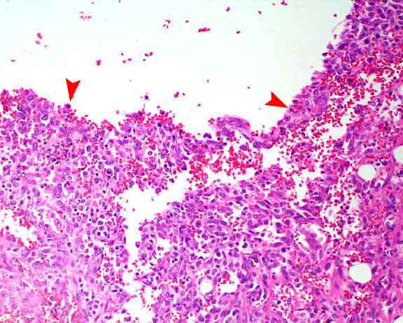
Another section of the tumour mass. Large, “cavernous”,
vascular spaces are filled with blood. (x40)
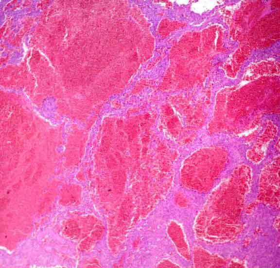
Vascular channels are lined by multiple layers of plump
endothelial cells. (Endothelial nuclei in haemangiomas and normal blood
vessels, are very flat). Mitotic rate is variable throughout the mass
(green arrows – mitotic figures). (x1000)
Haemangiosarcoma:
Haemangiosarcoma is the malignant variant of haemangioma
and is a common lesion in the spleen, heart, skin and subcutis of dogs.
Microscopically, the neoplastic endothelial cells of both haemangiomas
and haemangiosarcomas attempt to form blood-filled vascular channels although
this is more obvious in benign than in malignant tumours. Haemorrhage
is common to both tumours.
= mesenchymal tumour
|
|