|
|
|
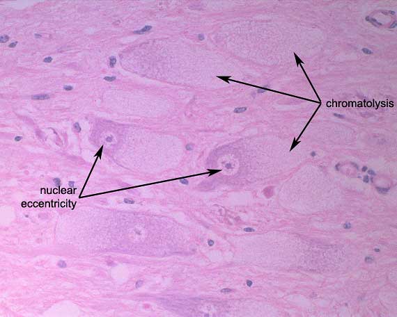
At high power, the cytoplasm
of the neuronal cell bodies appears foamy to finely vacuolated. This
appearance is very suggestive of a lysosomal storage disorder.
Assay of plasma a-mannosidase activity confirmed that
the steer had a-mannosidosis, an inherited lysosomal storage disorder
resulting from an inability to enzymatically degrade mannose-rich oligosaccharides
derived from glycoprotein.
The undegradable substrate accumulates within lysosomes
of neurons, glial cells, macrophages, fibroblasts, vascular endothelial
cells and epithelial cells in various organs. Storage in the permanent
neuronal cell population is responsible for the clinical signs.
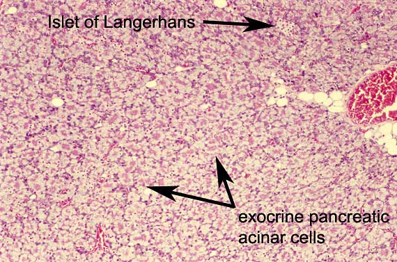
There is also marked vacuolation
of exocrine pancreatic acinar cell cytoplasm due to storage of oligosaccharides.
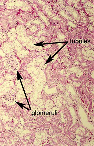
Severe storage is also apparent
within renal tubular epithelial cells.
a-mannosidosis was once common in Aberdeen Angus, Murray
Grey and Galloway cattle but has now largely been eradicated following
the development of assays to detect heterozygous carrier cattle. The latter
are phenotypically normal but have an approximately 50% reduction in the
plasma activity of a-mannosidase.
Identical histological neuronal lesions can be found
in ruminants poisoned by Swainsona species (e.g. Darling pea) in Australia
and by locoweeds (e.g. Astragalus and Oxytropis species) in North America.
These plants contain swainsonine, an indolizidine
alkaloid which inhibits lysosomal a–mannosidase.
|