Introduction
 |
||
 |
 |
|
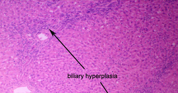
The increased prominence of the portal areas largely reflects biliary hyperplasia. The proliferating bile ductules/cholangioles are accompanied by sparse leukocytes (chiefly small lymphocytes) and by a mild excess of collagen. The latter in part reflects genuine fibrosis (portal or biliary fibrosis) but condensation of original connective tissue due to parenchymal atrophy is probably also contributing.
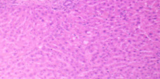
Have you found the other active area in the low power image?