|
|
|
Case 2:
History:
A 2 year old Abyssinian cat presented with a 3 week
long history of inappetence and lethargy. Physical examination revealed
mild pallor of the oral mucous membranes and palpable hepatomegaly. Haematology
and serum biochemistry revealed a mild regenerative anaemia and mild elevation
of AST, ALT, GGT, ALP and bilirubin.
Prior to surgery, prothrombin time (PT) and activated
partial thromboplastin time (APTT) assays were performed to assess for
any clotting factor deficiency attributable to liver dysfunction. The
PT and APTT were normal.
At exploratory laparatomy, the liver was diffusely
enlarged to a moderate degree, with rounding of all lobe borders. The
parenchyma was diffusely pale, with an exaggerated zonal pattern. The
parenchyma was also fragile and tore repeatedly during collection of a
peripheral wedge biopsy.
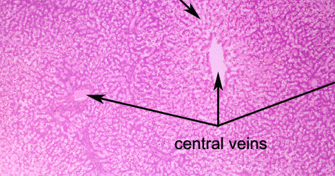
This is a low power view of the cat’s liver. Note
the mild undulation of the capsular surface. Central veins are dilated
and the radiating cords of hepatocytes appear very narrow, especially
in centrilobular zones.
PREVIOUS
|


