|
|
|
Case 4:
History:
Three 6 month old Merino weaners out of a flock of 500
died suddenly in good nutritional condition. The sheep were on dry pasture
and fed 0.75 kg of oats twice weekly. At necropsy of one of the sheep,
the liver was mildly swollen, mottled red-yellow, friable and tan discoloured,
with a patchy zonal pattern. Fresh fibrin strands were present over the
surface of the liver and between apposed liver lobes. There was mild oedema
of the gall bladder and hilar connective tissues, petechiation of the
gall bladder and a mild excess of clear yellow serous fluid in the peritoneal
cavity.
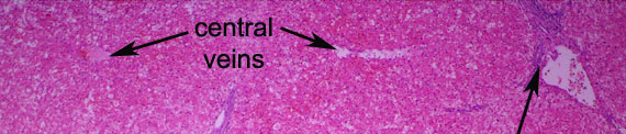
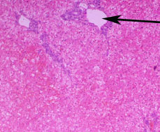
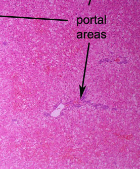
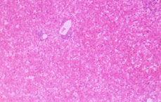
In this low power view of the liver, much of the parenchyma
appears redder than normal. A higher magnification will be needed to determine
if this is congestion versus haemorrhage and necrosis. Portal areas are
readily identifiable. Recognition of these is a useful first step in identifying
the precise nature of zonal patterns of hepatic injury.
PREVIOUS
|