Introduction
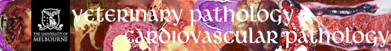
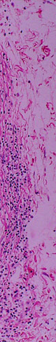
Blood vessels:
Most of the cells infiltrating the subintima and tunica
media of the artery are lymphocytes. The bright pink extracellular material
in the vessel wall (top right) is extravasated fibrin. The nuclei of intimal
endothelial cells (left) are irregularly swollen.
 |
 |
 |
||
| |
||||
 |
 |
|
||
| |
|
|
|
|
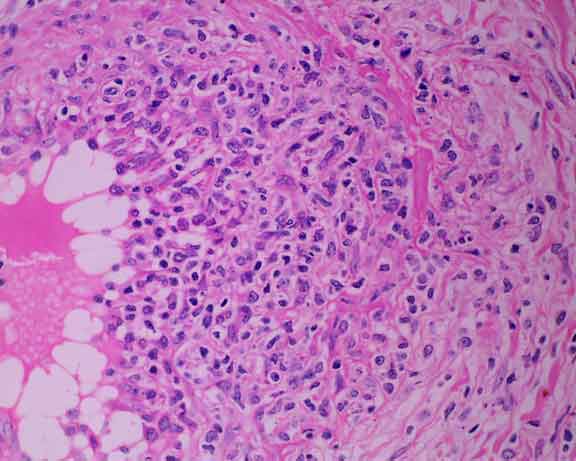
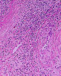
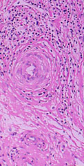
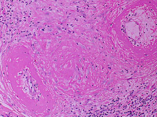
All of the arteries in this field are abnormal.