|
|
|
Blood vessels:
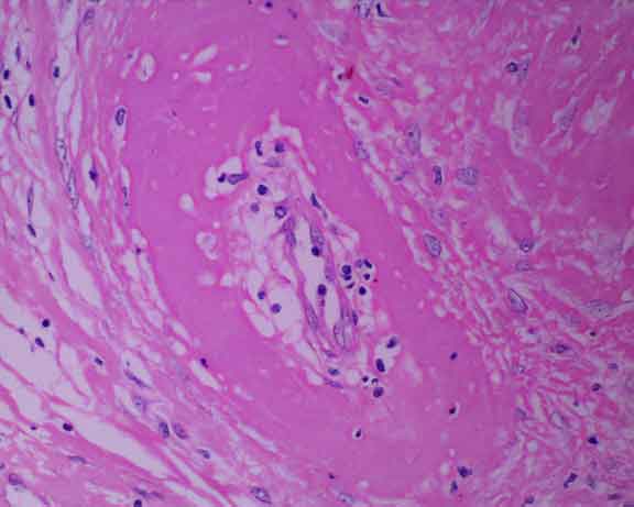
The vessel wall appears brightly eosinophilic due to
fibrin polymerisation in the areas of medial necrosis. The endothelium
of the intima is separated from the rest of the vessel wall by oedema
fluid. The still surviving endothelial cells have swollen nuclei. A few
leukocytes have infiltrated the damaged wall.
Fibrinoid necrosis is a
feature of many acute degenerative, necrotising and inflammatory disorders
of blood vessels. For example, it can be seen in vasculitis, renal failure,
hypertension, vitamin E or selenium deficiency and following vessel injury
by bacterial toxins.
|