
Introduction
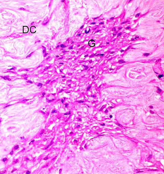
Haematoxylin & Eosin (x400)
Within the dermis, residual dermal collagen (DC) has a different histological appearance compared with granulation tissue (G) = fibroblasts, macrophages and new capillaries
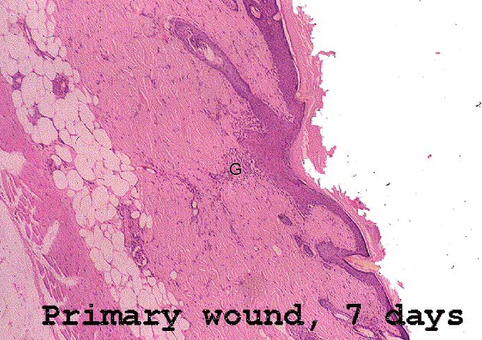
Haematoxylin & Eosin
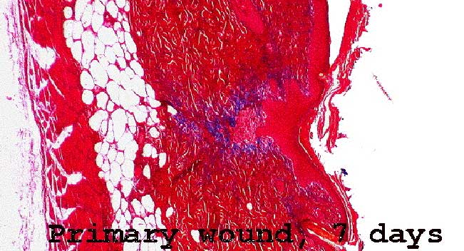
Trichrome stain
This stain highlights the immature collagen which is
being laid down at the wound edges, between the normal dermal collagen
and the granulation tissue of the wound. Newly deposited collagen stains
blue in this section.
![]()
![]()