The Patient
Unit 4: Preoperative Assessment & Management:
Topic 5: Surgical Planning
Careful surgical planning is an essential part of preoperative care for the patient.
In planning surgical procedures, either elective or in emergency situations, it is important to consider a number of factors:
- Risks vs. Potential benefits of the intended procedure
- Choice of technique to be used – based on experience and knowledge.
- Experience and technical skills of the surgeon. A good surgeon will always know their limitations.
- Facilities including adequate equipment and anaesthetic support. It is also important to be prepared for things that may go wrong!
- Client communication
- An understanding of the patients condition.
- An understanding of the surgical procedure.
- Prognosis, potential for complications
- Postoperative care needs
- Cost of the procedure, ability to pay.
The type of surgery to be performed may be classified in a number of ways:
- Diagnostic, curative, palliative and preventative.
- Emergency, non-elective and elective.
- Open, Endoscopic, Cryosurgery, Laser surgery.
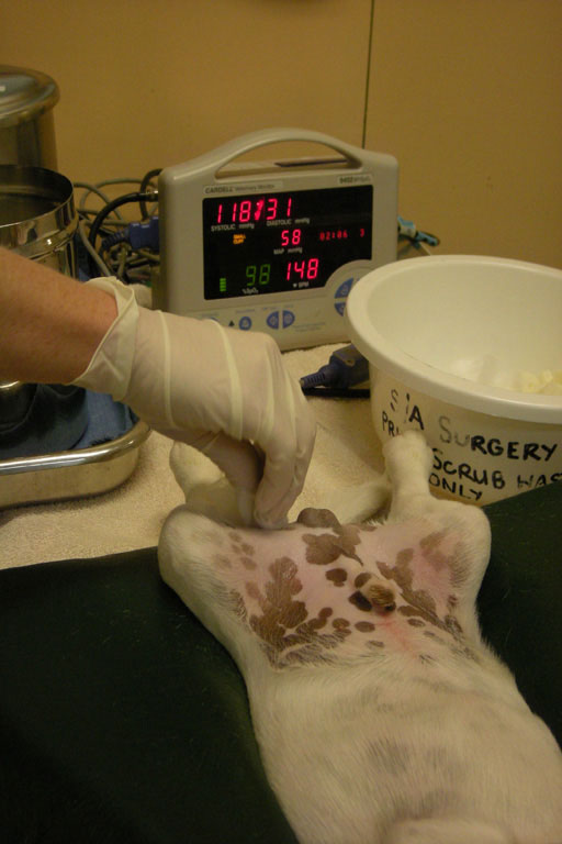
In planning for the surgical procedure and in recognising the potential for complications or for existing problems, the surgeon must work to optimise the conditions in which the procedure is undertaken in order to achieve a successful outcome.
In an emergency it is not always possible to completely stabilise the animal, but in some circumstances surgery may be needed quickly.
A number of methods may be used to reduce the risks of the anaesthetic and surgical procedure.
These include:
- Fluid therapy
- Blood Replacement
- Antibiotic Therapy
- Pre-emptive analgesia
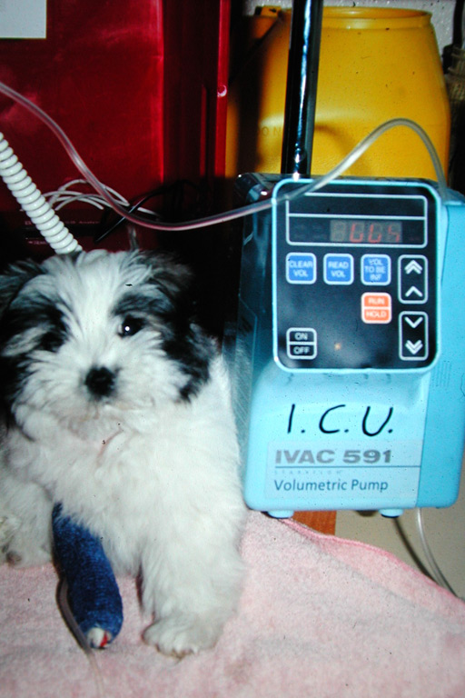
Fluid therapy
Even in emergency situations it is usually possible to start replacing fluid deficits and correcting acid-base and electrolyte imbalances.
Intravenous fluids are recommended for all significant surgical procedures, even in healthy animals. Unless given to correct a pre-existing deficit, they are normally administered at a maintenance rate of 10m/kg/hr.
Dehydration may result from a number of conditions associated with fluid loss including those associated with vomiting and iarrhea such as intestinal obstruction. The degree of dehydration can be assessed by the degree of skin turgor, sunken eyes, PCV and total protein, and can vary from 5% (mild), 10% (moderate) to (15%) severe. Treatment is by infusing a crystalloid fluid such as isotonic saline.
Fluid replacement (24 hour plan):
- 40-60 mL/kg/day plus continued loss in urine and faeces.
- Infuse half deficit in 20-30 minutes, but monitor PCV (>30%) and total protein (>3.5g/dL).
- Infuse other half plus maintenance, plus continued loss over the next 24 hours.
- Acid/base deficits may be estimated in association with the disease process, or more objectively with a blood gas analyser.
Fluid therapy – Acid/Base deficits
Respiratory acidosis
Usually results from depressed respiration or poor ventilation e.g. pneumothorax, pulmonary oedema.
- pH<7.35
- PCO2>35 torr
- HCO3>24 mEq/L
It is treated by correcting the cause.
Respiratory alkalosis
This may occur occasionally and usually results from hyperventilation, the most common cause being pain.
- pH>7.45
- pCO2<35 torr
- HCO3<20 mEq/L
It is treated by correcting the cause of the pain, or by administering analgesics.
Fluid therapy – Acid/Base deficits
Metabolic acidosis
This usually results from the loss of HCO3 due to diarrhoea or vomiting secondary to intestinal obstruction, or from the accumulation of acid such as in shock.
- pH<7.35
- HCO3<20 mEq/L
The body responds by increasing respiratory rate to try to eliminate the excess hydrogen ions.
It can be treated by administering Lactated Ringer’s solution.
In severe cases bicarbonate may be used, although its efficacy has been questioned recently.
MEq of HCO3 to be given = 0.3 X base deficit X B.Wt (kg)
The base deficit can be measured with a blood-gas analyser or estimated:
- Moderate – 5 mEq/L
- Marked – 10 mEq/L
- Severe – 15 mEq/L.
Give half intravenously over 10-15 minutes and re-evaluate. Give the remainder over four to six hours (if necessary).
Blood replacement
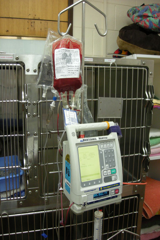
Blood transfusions are of benefit to animals with a PCV of less than 20%. The tranfused blood may help organ oxygenation, postoperative recovery rates and the healing process.
Contracted blood volume usually results in shock and may have a haemorrhagic, neurogenic or toxic origin. The amount of blood required can be calculated on the basis of the PCV required, that of the donor blood and the body weight of the animal.
The normal volume of blood in dogs and cats can be estimated on the basis of their body weight:
- 9% Body weight in dogs
- 7% Body weight in cats.
The PVC should be corrected to 30%, and total protein above 3.5g/dl.
Blood Volume Required = B.W. (Kg) x Desired PCV - Patient PCV x 90*
Donor PCV
* 70 for cats
Treating Pre-existing Coagulation Deficits
The use of whole blood should be avoided in the treatment of coagulopathies due to the risk of future transfusion reactions if an un-crossmatched transfusion is subsequently given.
Haemophilia A is an inherited, sex-linked deficiency of factor VIII seen mostly in male German Shepherd dogs. The treatment of choice is cryoprecipitate or fresh frozen plasma as a single dose, or as an infusion during surgery.
Von Willebrand disease is prevalent in Dobermans and Scottish Terriers in Australia. Up to 60 per cent of Dobermans tested have this disorder. The treatment and prevention of haemorrhage during surgery is the same as for haemophilia A.
Disseminated intravascular coagulation may be a serious complication of a number of conditions including haemangiosarcoma, heat stroke, gastric-dilation volvulus, pancreatitis, liver disease and sepsis. DIC contributes significantly to patient morbidity and mortality. Therapy of DIC relies primarily upon correction of the underlying disorder. Replacement of coagulation factors and antithrombin III may be achieved with a plasma infusion. Whole blood is indicated in severe hrombocytopenia or severe haemorrhagic anaemia.
Chronic liver disease may result in reduced vitamin K-dependent coagulation factors and albumin. Plasma therapy can be useful if there are clinical signs of haemorrhage.
The dose rate of plasma is 6–10 mL/kg given two to three times daily until haemorrhage ceases. It should be given slowly through a separate catheter and infusion set to Hartmann’s solution as there is enough ionised calcium in Hartmann’s to initiate clotting in the plasma.
Preoperative antibiotic therapy
The use of preoperative antibiotics is generally dictated by either the disease process or the surgical procedure to be undertaken.
They should always be used in the face of an ongoing infection, and should be given to clear or control the infection prior to the surgical procedure if possible.
Antibiotics are not usually required for routine aseptic surgery, however for areas in which the development of an infection would be disastrous, such as the nervous system or joints, antibiotics are frequently used prophylactically prior to the procedure.
There is also a role for preoperative antibiotics in areas of the body where complete avoidance of contamination is not always possible, such as surgery on the large bowel. This is further discussed in the later modules dealing with specific areas of the body.
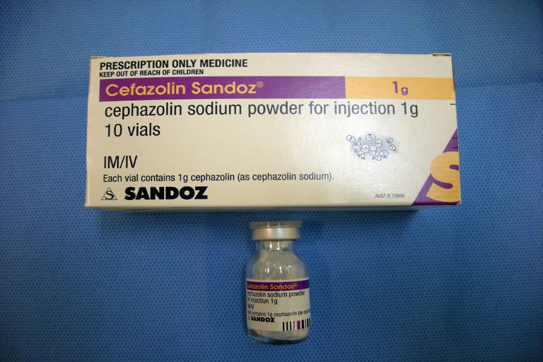
The aim of antibiotic prophylaxis is to achieve a high tissue level of the antibiotic during the procedure. In most cases this requires the use of intravenous antibiotics at induction.
We routinely use Cephazolin (20mg/kg) intravenously at induction and repeat the dose following two hours of surgery, or 8 hours postoperatively.

Howe, L.M., & Boothe, H.W. (2006) Antimicrobial use in the surgical patient. Vet Clin Small Anim 36: 1049-1060.